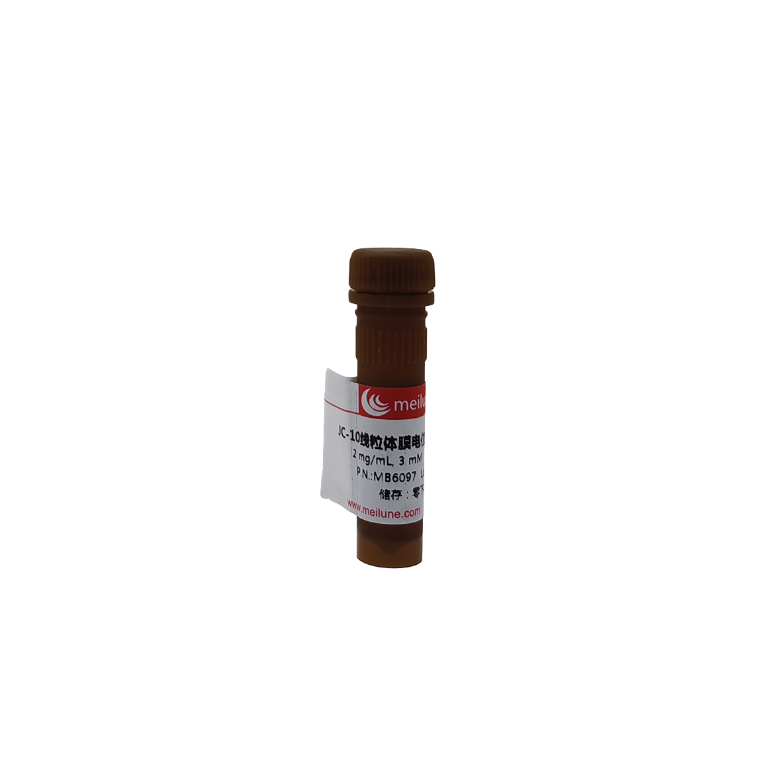
JC-10线粒体膜电位荧光探针溶液

¥1,180.00

规格: 5X100ul

英文名字:JC-10 ;JC-1的卓越代替品*
质量标准:溶液2 mg/mL, 3 mM
货号: MB6097-1

JC-10线粒体膜电位荧光探针溶液;JC-10 ;JC-1的卓越代替品*

虽然JC-1在许多实验中被广泛地的实验,但是其水溶性差的特性也使其很难在一些实验中使用。即使是在浓度为1 uM的条件下,JC-1也会在水的缓冲液中析出。JC-10是开发的一种可以在需要高浓度染料的实验中替代JC-1。与JC-1相比,我们的JC-10具有更好的水溶性。JC-10可以选择性的进入线粒体,并且由于膜电位的增加,其颜色会发生从绿色到橙色的可逆性改变。这种特性是由于JC-10聚合物的可逆结构,在膜极化条件下,它的发射光由520nm(JC-10 单体发射光)转移到570nm (J-聚合体发射光)。当在490nm处激发时,JC-10发生从绿色到橙绿色的可逆改变,因为线粒体膜极化的更大。这两种颜色都可以被流式细胞仪上装好的一般滤光器检测到,因此绿色发射光可以通过荧光通道1(FL1)进行分析,橙绿色发射光可以通过荧光通道2(FL2)进行分析。它除了有潜力用于流式细胞术,也可以用于荧光成像分析。We have developed a protocol to use it in fluorescence microplate platform.。在一些细胞系中JC-10有着比JC-1更好的表现;有趣地是,JC-10的性能表现对细胞系极具依赖性。我们的所提供的JC-10溶解于DMSO溶液中,其浓度为3mM (2 mg/mL).

规格

5×100 uL

产品形式

3mM(2mg/ml) 溶液

Ex (nm)

510

Em (nm)

525

分子量

~600

溶剂

DMSO

1. Prepare JC-10 working solution:
1.1 Each vial of DMSO stock solution (100 µL, 2 mg/mL, 3 mM) should be used only once. Any unused vials should be stored at<-20℃.
Note: Avoid repeated freeze-thaw cycles, and protect from light.
1.2 Prepare a 1X JC-10 working solution: On the day of the experiment, thaw an aliquot of the JC-10 stock solution to room temperature. Prepare a 10 to 30 µM 1X working solution in Hanks and 20 mM Hepes buffer (HHBS) or buffer of your choice, pH 7-8 with 0.02% Pluronic® F-127. Mix them well by votexing.
Note: For some cell lines, working solution at pH 8 might prevent JC-10 leakage.
2. Run JC-10 assay with a fluorescence microplate reader:
2.1 Treat cells with test compounds for a desired period of time (For example, Jurkat cells can be treated with camptothecin for 4-6 hours) to induce apoptosis. For blank wells (medium without the cells), add the corresponding amount of compound buffer.
2.2 Add 100 µL/well/96-well plate or 25 µL/well/384-well plate of JC-10 working solution (from Step 1.2) into the cell plate.
2.3 Incubate the JC-10 loading plate in a 37℃, 5% CO2 incubator for 15-60 min.
Note: The appropriate incubation time depends on the individual cell type and cell concentration used. Optimize the incubation time for each experiment.
2.4 Monitor the fluorescence change at Ex/Em = 490/525 nm (FITC channel) and 540/595 nm (TRITC channel) for ratio analysis.
Optional: Remove the JC-10 working solution from the plate; add 100 µL/well/96-well plate or 25 µL/well/384-well plate of HHBS back to the cell plate before analysis.
3. Run JC-10 assay with a fluorescence microscope or a flow cytometer:
3.1 Treat cells with test compounds for a desired period of time (For example, Jurkat cells can be treated with camptothecin for 4-6 hours) to induce apoptosis.
3.2 Centrifuge the cells to get 1-5 x 10⁵ cells per tube.
3.3 Resuspend cells in 500 µL of JC-10 working solution (from Step 1.2).
3.4 Incubate at room temperature or in a 37 °C, 5%CO2 incubator for 10 to 30 min, protected from light.
Note: The appropriate incubation time depends on the individual cell type and cell concentration used. Optimize the incubation time for each experiment.
3.5 Monitor the fluorescence change at Ex/Em = 490/525 nm and 540/595 nm with a fluorescence microscope (using FITC and TRITC filters) or a flow cytometer (using FL1 and FL2 channels).
Optional: Remove the JC-10 working solution from the plate; add 100 µL/well/96-well plate or 25 µL/well/384-well plate of HHBS back to the cell plate before analysis on fluorescence microscope.

运输条件:2~8℃运输

相关产品推荐

MA0061

Pluronic™ F-127 (20% Solution in DMSO)

MB6055

JC-1线粒体膜电位荧光探针

备注:所有报价均已含国内快递费及发票费用。库存情况可能有所变动,请与我们销售人员确认为准,价格情况请直接联系我们销售人员。

我司所售出产品仅供于科研研究用途(非临床科研研究),每次销售产品行为都适用于我司网上所列明的通用销售条款

规格

5X100ul

相关文档

购物车
滚动至顶部