
产品简介:
EGTA是最为被广泛使用的钙离子选择性的螯合剂。它与钙离子形成的复合物比与镁离子形成的复合物要稳定100,000倍。它被用来制备钙缓冲液和控制钙离子的浓度。EGTA, AM是EGTA的乙酰甲酯衍生物,通过AM法,能被轻易负载到细胞中。
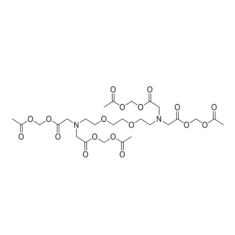
分子式:C26H40N2O18;分子量:668.6
溶解性:溶于DMSO
储存条件:
-20℃
- 使用说明(仅供参考)
- 1.Load Cells with Calcium Indicator AM Esters:
AM esters are the non-polar esters that readily cross live cell membranes, and rapidly hydrolyzed by cellular esterases inside live cells. AM esters are widely used for loading a variety of polar fluorescent probes into live cell non-invasively. However, cautions must be excised when AM esters are used since they are susceptible to hydrolysis, particularly in solution. They should be reconstituted in high-quality, anhydrous dimethylsulfoxide (DMSO). DMSO stock solutions should be stored desiccated at -20 °C and protected from light. Under these conditions, AM esters should be stable for several months.
Following is our recommended protocol for loading AM esters into live cells. This protocol only provides a guideline, and should be modified according to your specific needs.- Prepare a 2 to 5 mM AM esters stock solution in high-quality, anhydrous DMSO.
- On the day of the experiment, either dissolve calcium indicators solid in DMSO or thaw an aliquot of the indicator stock solutions to room temperature. Prepare a working solution of 2 to 20 µM in the buffer of your choice (such as Hanks and Hepes buffer) with 0.04%Pluronic® F-127. For most cell lines we recommend the final concentration of calcium indicators be 4-5 uM. The exact concentration of indicators required for cell loading must be determined empirically. To avoid any artifacts caused by overloading and potential dye toxicity, it is recommended to use the minimal probe concentration that can yield sufficient signal strength.
- If your cells (such as CHO cells) containing the organic anion-transports, probenecid (2–5 mM) or sulfinpyrazone (0.2–0.5 mM) may be added to the the dye working solution (final in well concentration will be 1-2.5 mM for probenecid, or 0.1 -0.25 mM for sulfinpyrazone) to reduce the leakage of the de-esterified indicators.
- Add equal volume of the dye working solution (from Step b or c) into your cell plate.
- Incubate the dye-loading plate room at temperature or 37 °C for 20 minutes (especially Fluo-8 AM) to 2 hours, and then incubate the plate at room temperature for another 30 minutes.
Note1: Decreasing the loading temperature might reduce the compartmentalization of the indictor.
Note1: Incubate the Cal-520 AM longer than 2 hours gives better signal intensity for some cell lines. - Replace the dye working solution with HHBS or buffer of your choice (containing an anion transporter inhibitor, such as 1 mM probenecid, if applicable) to remove excess probes.
- Run the experiments at desired Ex/Em wavelengths (see Table 1).
2.Measure Intracellular Calcium Responses:
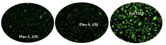
Figure 1.Response of endogenous P2Y receptor to ATP in CHO-M1 cells withoutprobenecid.CHO-M1 cells were seeded overnight at 40,000 cells per 100 µL per well in a 96-well black wall/clear bottom costar plate. 100 µl of 4 µM Fluo-3 AM, Fluo-4 AM or Cal 520® AM in HHBS were added into the wells, and the cells were incubated at 37 °C for 2 hour. The dye loading medium were replaced with 100 µl HHBS, 50 µl of 300 µM ATP were added, and then imaged with a fluorescence microscope (Olympus IX71) using FITC channel.
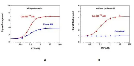
Figure 2.ATP-stimulated calcium response of endogenous P2Y receptor in CHO-K1 cells measured with Cal-520® or Fluo-4 AM.CHO-K1cells were seeded overnight in 50,000 cells per 100 µL per well in a 96-well black wall/clear bottom costar plate. 100 µL of 5 µM Fluo-4 AM or the Cal-520® AM with (A) or without (B) 2.5 mM probenecid was added into the cells, and the cells were incubated at 37oC for 2 hours. ATP (50µL/well) was added by FlexStation (Molecular Devices) to achieve the final indicated concentrations.